Particle Size Analysis in Oral Thin Films using Optical Microscopy and CLAIRITY™ PARTICLE

Introduction

Overview

Oral thin films contain an active pharmaceutical ingredient (API) embedded within a polymeric matrix. Evaluating particle size directly in the film can help formulation teams understand particle distribution while preserving the particles’ in-film state. This case study outlines how optical microscopy combined with the CLAIRITY™ PARTICLE Module can support particle characterization during formulation development and provide visual evidence relevant to regulatory justification.

The Analytical Challenge

In oral thin films, the API is dispersed within a polymeric matrix. The analytical workflow therefore needs to visualize embedded particles without unnecessarily disturbing their native state, while helping analysts assess particle size, agglomeration, and dispersion uniformity. A workflow based on microscopy and image analysis offers direct visual context alongside quantitative measurements

The Approach

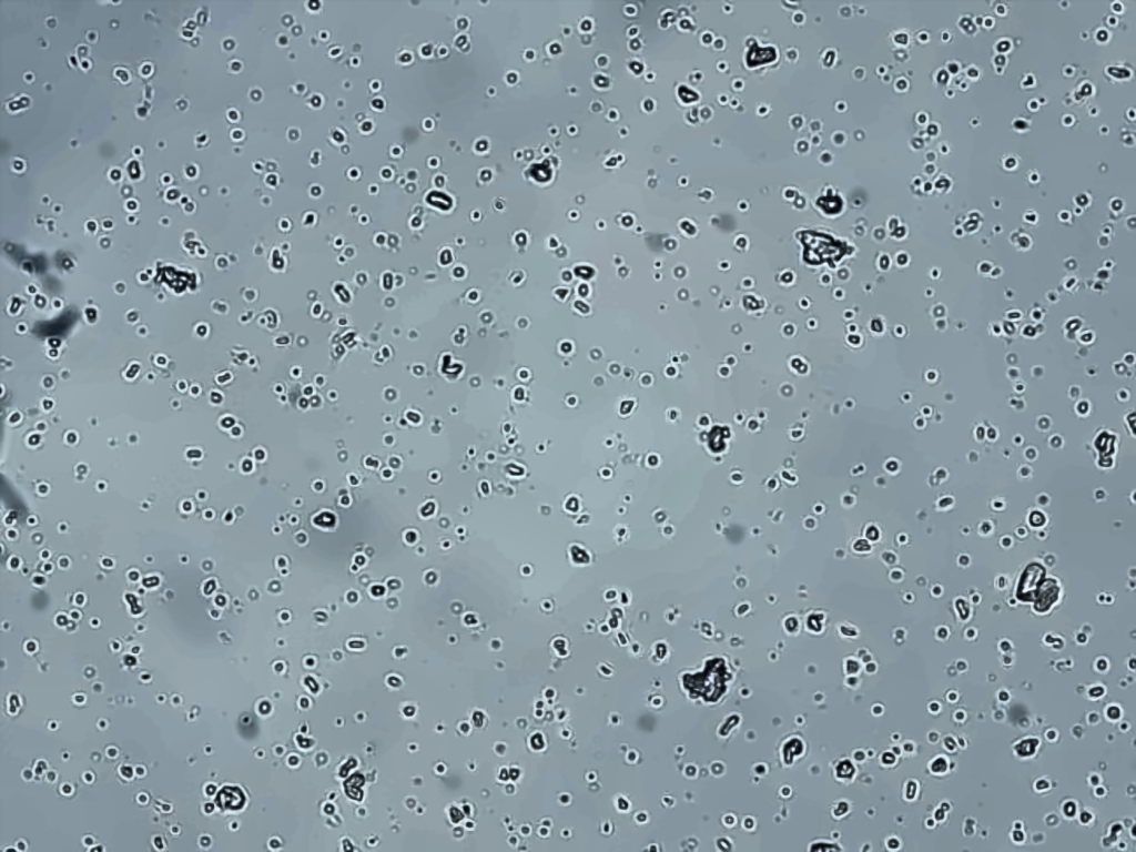
With careful sample preparation and upright optical microscopy, embedded API particles can be visualized directly within the film. Images can then be evaluated using the CLAIRITY™ PARTICLE Module to turn microscopic observations into structured particle-analysis data.

Workflow

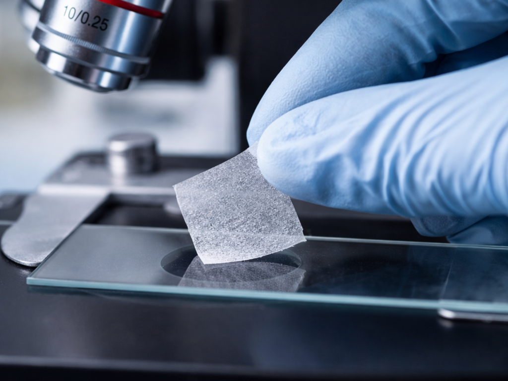
  • Prepare the sample — Prepare the oral thin film carefully to support visualization of embedded particles while maintaining their in-film state.
  • Capture microscopic images — Use upright optical microscopy to observe particles within the film.
  • Detect and classify particles — Apply the CLAIRITY™ PARTICLE Module for automated particle detection and classification.
  • Review size distribution — Generate particle-size distribution metrics, including D10, D50, and D90.
  • Assess dispersion — Review particle counts, agglomeration, and dispersion uniformity, using image evidence to support interpretation

Capabilities of the CLAIRITY™ PARTICLE Module

  • Automated particle detection and classification
  • Particle-size distribution reporting (D10, D50, D90)
  • High-throughput particle counting
  • Assessment of agglomeration and dispersion uniformity
  • Support for differentiating API particles from the polymer matrix

Analytical Value

This workflow connects microscopic observations with quantitative, reproducible particle-analysis data. It can support formulation-development decisions by helping teams evaluate particle size and distribution, identify potential agglomeration, and review dispersion uniformity. The combination of image evidence and quantitative outputs can also contribute to documentation prepared for regulatory justification.

Conclusion

Optical microscopy paired with the CLAIRITY™ PARTICLE Module provides a practical approach for assessing API particles directly within oral thin films. By combining automated detection and classification with particle-size distribution metrics and visual review, the workflow supports a more structured understanding of particle characteristics during product development.