Talc is extensively used in pharmaceutical, cosmetic, and personal care formulations, where the presence of asbestos poses serious health and regulatory concerns. Regulatory authorities therefore require stringent monitoring and reliable identification of asbestos contamination.
Consequently, there is an increasing demand for a practical, microscopy-based screening and evaluation approach that is reliable, reproducible, and cost-effective for routine laboratory use.
This study was conducted by using ImageProVision CLAIRITYTM – PARTICLE System and Polarized Microscope Configuration as per USP recommendation.
The objective of this research at ImageProVision is to:
• Develop and validate a microscopy-based method for asbestos identification in talc
• Perform qualitative and quantitative analysis based on optical and birefringence characteristics
• Demonstrate that the CLAIRITYTM – PARTICLE system provides a robust alternative to high-end analytical techniques for routine asbestos analysis
A. Wet Sieving Concentration.
1. Sieve Cleaning:
• Use 76-mm (3-inch), 400-mesh (37 μm) sieve.
• Clean in detergent solution using sonic bath for 15 minutes.
• Rinse thoroughly with deionized water.
2. Sample Weighing:
• Weigh approximately 2 g of talc powder into a 100-mL plastic bottle with lid.
3. Sample Wetting & Dispersion:
• Wet completely with isopropyl alcohol (IPA).
• Add deionized water to ~75 mL (¾ full).
• Shake vigorously for 30 seconds.
4. Sieving:
• Slowly pour suspension onto 400-mesh sieve.
• Discard passing fraction as per local regulations.
•Rinse bottle repeatedly with water (and IPA if foaming), shake, and pour until
bottle is clean.
5. Sieve Rinsing:
• Rinse gently with water spray to avoid splashback.
• Continue until water passing through is clear and mesh is not blinded.
• A small visible residue should remain on the sieve.
6. Residue Collection & Drying:
• Rinse retained residue into a tared aluminium pan or glass dish using particlefree water.
• Dry at room temperature or at 105–110°C.
• Weigh dried residue.
7. Residue Requirement:
• Target: 0.05–0.1 g residue for PLM analysis.
• If <0.05 g obtained:
• Repeat procedure, or
• Use larger starting quantity, or
• Use finer mesh sieve.
B. Sample Preparation of Sieved Residue (For PLM)
8. Slide Loading:
• Place known quantity of residue on clean glass slide.
• Ensure 30–50% area coverage under coverslip.
9. RI Liquid Addition:
• Add 1–2 drops of appropriate refractive index (RI) liquid.
• Disperse particles evenly using clean metal probe.
10. Coverslip Placement:
• Place coverslip gently.
• Apply light pressure using soft-tipped tool to remove air bubbles and improve dispersion.
11. Minimum Sample Requirement:
• Analyse minimum 5 mg total per RI liquid.
• Typically prepare 3–5 slides (1–2 mg each) as required for PLM asbestos identification.
1. CLAIRITYTM – PARTICLE Module Software with High end Polarised Trinocular microscope, equipped with rotating stage.
2. IPV High Resolution Camera (5 which has 2560 X 1920 Resolution also CMOS – Sony IMX335LQN – C sensor.
3. 1.550 and 1.605 RI liquids
4. 76-mm (3-inch), 400-mesh (37 μm) sieve.
5. 0.45 μm membrane filter paper
6. Glass slides & coverslips
• Asbestos identification is performed using the dispersion staining technique under a polarized light microscope (PLM).
• The microscope is configured in plane-polarized light (PPL) mode, without condenser, and equipped with a centered dispersion staining objective.
• Slides are prepared using refractive index (RI) liquids of 1.550 and 1.605.
• Each slide is scanned completely in an orthogonal pattern using a mechanical stage.
• The slide is then rotated 90° and rescanned to minimize the risk of missing fibers.
• Suspect fibers are identified based on characteristic dispersion staining colors:
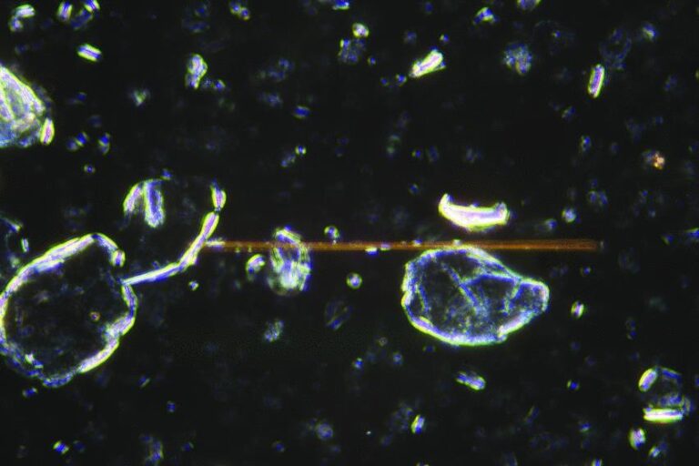
• Chrysotile: magenta (nǁ) and blue (n⊥) in RI 1.550.
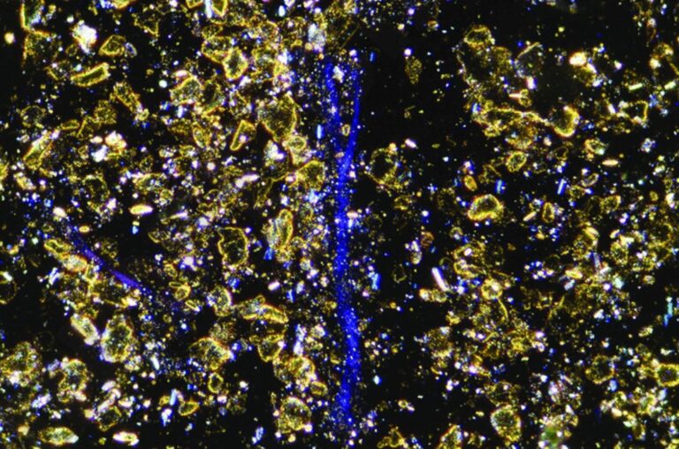
• Amphiboles: yellow (nǁ) in RI 1.605, with variable perpendicular colors.
• Refractive indices are confirmed using the Becke line technique.
• Amphibole RI is typically >1.605.
• Talc RI is typically <1.605.
• Optical properties documented at 100×–400× magnification include:
• Morphology
• Aspect ratio
• Pleochroism
• Extinction characteristics
• Sign of elongation
• Birefringence
• RI behavior
• Fibers are evaluated for asbestiform characteristics such as:
• High aspect ratio (≥20:1 for fibers >5 µm)
• Thin fibrils (<0.5 µm)
• Parallel bundles
• Splayed ends
• Curvature or matted masses
• All confirmed suspect fibers are digitally documented.
• The working detection limit is 0.01% w/w (100 ppm).
• A minimum of two confirmed asbestos fibers is required to report a positive result.
• Zero or one confirmed fiber is reported as below detection limit (<0.01% w/w or <100 ppm).
Representative polarized light micrographs illustrating asbestos fiber identification and differentiation from talc particles under various imaging conditions.